The macula is an area of the central retina which is most densely packed with retinal light receptor cells known as cones. The macula is the part of the retina which allows for fine central used in reading, facial recognition and performing fine motor tasks.
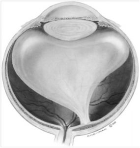
The back chamber of the eye is filled with a jelly-like substance called the vitreous. The vitreous is fairly well formed or solid in early life, but with age, fluid filled pockets form within the substance of the vitreous. As this occurs, the vitreous collapses in on itself and separates or detaches from the surface of the retina, in a process known as a posterior vitreous detachment. When this process is incomplete, the vitreous may be stuck on to the retina in certain areas. These areas are known as abnormal vitreoretinal interface abnormalities. In the peripheral retina, this incomplete separation of the vitreous from the retina and ongoing traction can result in a retinal tear. At the macula, an abnormally strong vitreomacular adhesion can result in a round hole formed at the centre of the macula, called a macular hole.
Figure 1: Abnormal vitreous adhesion to the macula, a precursor to macular hole formation
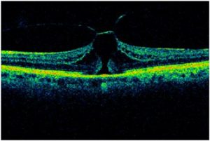
Figure 2 : – Optical Coherence Tomography (OCT) scan of a Pre – full thickness macular hole
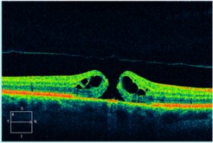
Figure 3 – Optical Coherence Tomography (OCT) scan of a full thickness macular hole
Macular hole surgery involves performing a vitrectomy whereby the vitreous is removed via small key holes to allow for instrumentation into the eye. Following the removal of the vitreous, a fine layer called the internal limiting membrane is peeled from the surface of the retina around the macular hole. The eye is then filled with a bubble of gas that takes approximately 3 weeks to be completely reabsorbed and replaced by the aqueous humour which is a saline like solution that the eye produces.
This bubble of gas changes the focal plane of light that enters the eye and as a result with a bubble of gas in the eye, one’s vision will be very blurred. Whilst there is a bubble of gas in the eye, due to the blurred vision caused by the gas, you will not be able to drive. Flying is not permitted as the gas bubble expands resulting in raised intraocular pressure which can lead to irreversible blindness.
Posturing in a face down position may be required following macular hole surgery and the need to do so will be discussed with you by your surgeon. The success rate for macular hole surgery is high with a greater than 90% chance of successful closure of the macular hole with a single operation. The improvement of vision may take time but is usually apparent once the gas bubble disappears. As with other operations involving vitrectomy and the injection of a gas bubble into the eye, patients may develop a cataract in the eye necessitating cataract surgery anytime from 3 to 12 months after macular hole surgery. It is important to realize that only after cataract surgery will one’s best visual potential in that eye be realized.
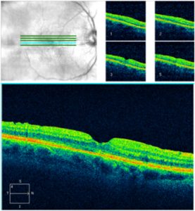
Figure 4: – Optical Coherence Tomography (OCT) scan showing a closed macular hole following surgery


