The back chamber of the eye is filled with a jelly-like substance called the vitreous. The vitreous is fairly well formed or solid in early life, but with age, fluid filled pockets form within the substance of the vitreous. As this occurs, the vitreous collapses in on itself and separates or detaches from the surface of the retina, in a process known as a posterior vitreous detachment. A posterior vitreous detachment may cause microscopic damage to the surface of the central retina or macula. The healing response initiated by the retina to repair the microscopic damage results in a superficial scar or membrane that grows over the surface to the macula like a skin. This is called an epiretinal membrane. A posterior vitreous detachment may also cause a tear in the retina, allowing pigment epithelial cells to migrate onto the surface of the retina further contributing to the formation of an epiretinal membrane.
Generally, the healing response is relatively mild and only results in a very thin layer of scar tissue growing across the surface of the macula thus causing minimal symptoms to the patient. However, some may have a thicker opaque layer of scar tissue forming over the surface of the retina which causes the central retina to have a rippled or puckered appearance. These patients may be symptomatic of distortion in their vision and may also have difficulties with both distance and reading vision. They may find that they are more comfortable closing the eye with the epiretinal membrane whilst reading due to image rivalry. For most people that have a significant epiretinal membrane, the growth is very slow and eventually stops progressing.
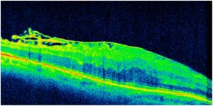
Figure showing an Optical Coherence Tomography (OCT) scan of the macula with a thick epiretinal membrane on the surface of the macula.
Surgery for Epiretinal Membrane
Treatment for epiretinal membranes is only necessary if the epiretinal membrane causes symptoms or is associated with a reduction in visual acuity. Epiretinal membrane surgery involves removal of the vitreous (vitrectomy surgery) which then allows us to grasp the membrane with a pair of fine forceps and gently peel it off the surface of the retina. At the time of epiretinal membrane surgery, the peripheral retina is also examined for retinal tears and any tears found will be treated with cryotherapy (freezing) performed to the retinal tear. This is performed to prevent the retina from detaching due to the retinal tear. As with retinal detachment surgery, a gas bubble may be used to splint the retina in place whilst the retinal tear that has been treated heals up. Epiretinal membrane surgery is very effective in alleviating the symptoms of distortion that one experiences due to the membrane puckering up the macula but the recovery of vision may take several months for maximal recovery.
The vitrectomy component of the surgery will accelerate cataract formation and it is important to realize that as the vision improves from the epiretinal membrane surgery itself, at some point, it may start to deteriorate from the cataract. Therefore, in patients who have not previously had cataract surgery prior to epiretinal membrane surgery the best recovery of vision will only be realized after cataract surgery.

