The eye can best be described as a complex organ with many parts. Good vision is dependent upon each part functioning as a whole.
When light enters the eye, it passes through the tear film, which coats the cornea. The cornea covers the front of the eye and assists in focusing incoming light.
The colored part of the eye is the iris. The iris widens to make the pupil bigger and constricts to make it smaller, as light conditions change. This enables more or less light to enter the eye.
Light next passes through the lens, a flexible and transparent part of the eye that can change its shape to focus images on the retina.
Next light passes through the center of the eye, known as the vitreous. The vitreous is filled with a clear jelly substance.
Lastly, light passes through the retina, which is a light-sensitive tissue that lines the back of the eye. The retina converts light into information that the brain can use.
The macula is in the central part of the retina. It contains a dense population of light sensing cells known as photoreceptors. The fovea is the center of the macula, and it contains the highest density of photoreceptors. The fovea enables us to see fine details, like small newsprint.
Behind the retina is the choroid, which is a layer of blood vessels that supply nutrients and oxygen to the outer layers of the retina. The optic nerve is a bundle of nerve fibers responsible for carrying visual information to the brain.

The Retina
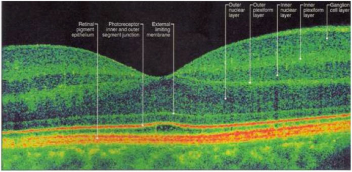
The retina is made up of many different layers of tissue, each responsible for a specific function. In patients with macular degeneration, some of these layers do not function as normal.
The photoreceptor layer is made up of rods and cones, which are light-sensitive cells. Inside these photoreceptors, light images are converted into electrochemical signals.
Under the photoreceptors is the retinal pigment epithelium or RPE. The cells of RPE are responsible for absorbing excess light and carrying nutrients, oxygen and other cellular wastes between the choroid and photoreceptors.
The blood vessels of the RPE layer and choroid are separated by Bruch’s membrane. The fibrous, white outer covering of the eye is known as the sclera.